Color blindness, also known as color vision deficiency, is the decreased ability to see color or differences in color. The most common cause of color blindness is an inherited fault in the development of one or more of the three sets of color sensing cones in the eye. Males are more likely to be color blind than females, as the genes responsible for the most common forms of color blindness are on the X chromosome. As females have two X chromosomes, a defect in one is typically compensated for by the other, while males only have one X chromosome.
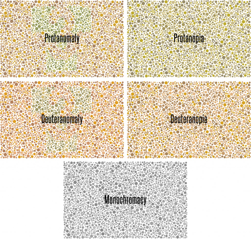
Monochromacy, also known as “total color blindness”, is the lack of ability to distinguish colors (and thus the person views everything as if it were on a black and white television); caused by cone defect or absence. Monochromacy occurs when two or all three of the cone pigments are missing and color and lightness vision is reduced to one dimension.
Protanomaly is a mild color vision defect in which an altered spectral sensitivity of red retinal receptors (closer to green receptor response) results in poor red–green hue discrimination. It is hereditary, sex-linked, and present in 1% of males.
Deuteranomaly, caused by a similar shift in the green retinal receptors, is by far the most common type of color vision deficiency, mildly affecting red–green hue discrimination in 5% of European males. It is hereditary and sex-linked.
Protanopia is a severe type of color vision deficiency caused by the complete absence of red retinal photoreceptors. Protans have difficulties distinguishing between blue and green colors and also between red and green colors. It is hereditary, sex-linked, and present in 1% of males.
Deuteranopia is a severe type of color vision deficiency where the green photoreceptors are absent. It affects hue discrimination in the same way as protanopia. Like protanopia, it is hereditary, sex-linked, and found in about 1% of the male population.
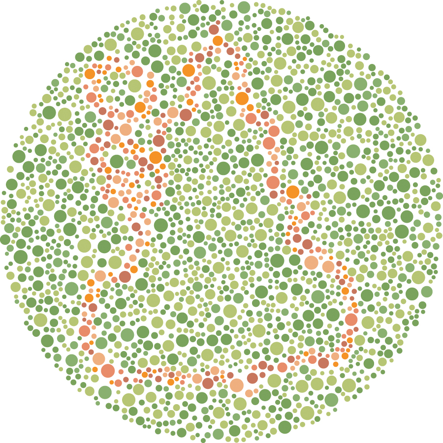
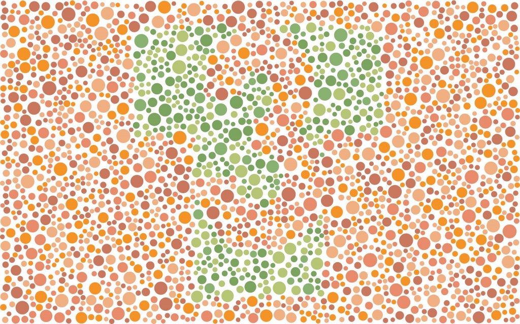
The Ishihara test is a color perception test for red-green color deficiencies, the first in a class of successful color vision tests called pseudo-isochromatic plates (“PIP”). It was named after its designer, Dr. Shinobu Ishihara, a professor at the University of Tokyo, who first published his tests in 1917
The test consists of a number of colored plates, called Ishihara plates, each of which contains a circle of dots appearing randomized in color and size. Within the pattern are dots which form a number or shape clearly visible to those with normal color vision, and invisible, or difficult to see, to those with a red-green color vision defect. Other plates are intentionally designed to reveal numbers only to those with a red/green color vision deficiency, and be invisible to those with normal red/green color vision. The full test consists of 38 plates, but the existence of a severe deficiency is usually apparent after only a few plates.
I thought, “why not make a T3 Ishihara Plate?” So I did. I found an online tool to help people see what an image looks like to someone with color vision deficiency and produced the following images:
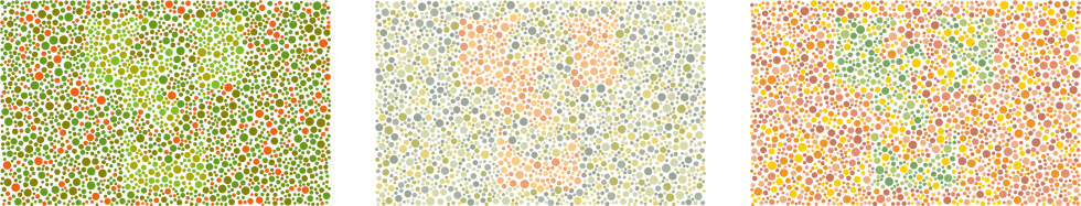
When I showed the T3 PIP plates to one of my graphic designers who is also our Curator, she asks if I was going to do a AAA. Hadn’t really thought about it, but why not…